Anatomy of the nose
Introduction to the anatomy of the nose
Wow!!! what a great smell. We can smell all the things because of our noses. Let’s dive into knowing more about the nose by learning the anatomy of the nose and nasal bones.
The human nose is the body’s primary organ for smell and also functions as part of the body’s respiratory system.
The nose anatomy bears the nostrils. It is also known as the chief organ in the olfactory system. The shape of the nose is obstinate by the nasal bones and the nasal cartilages, which are discussed below in the main article.
Anatomy of the External nose
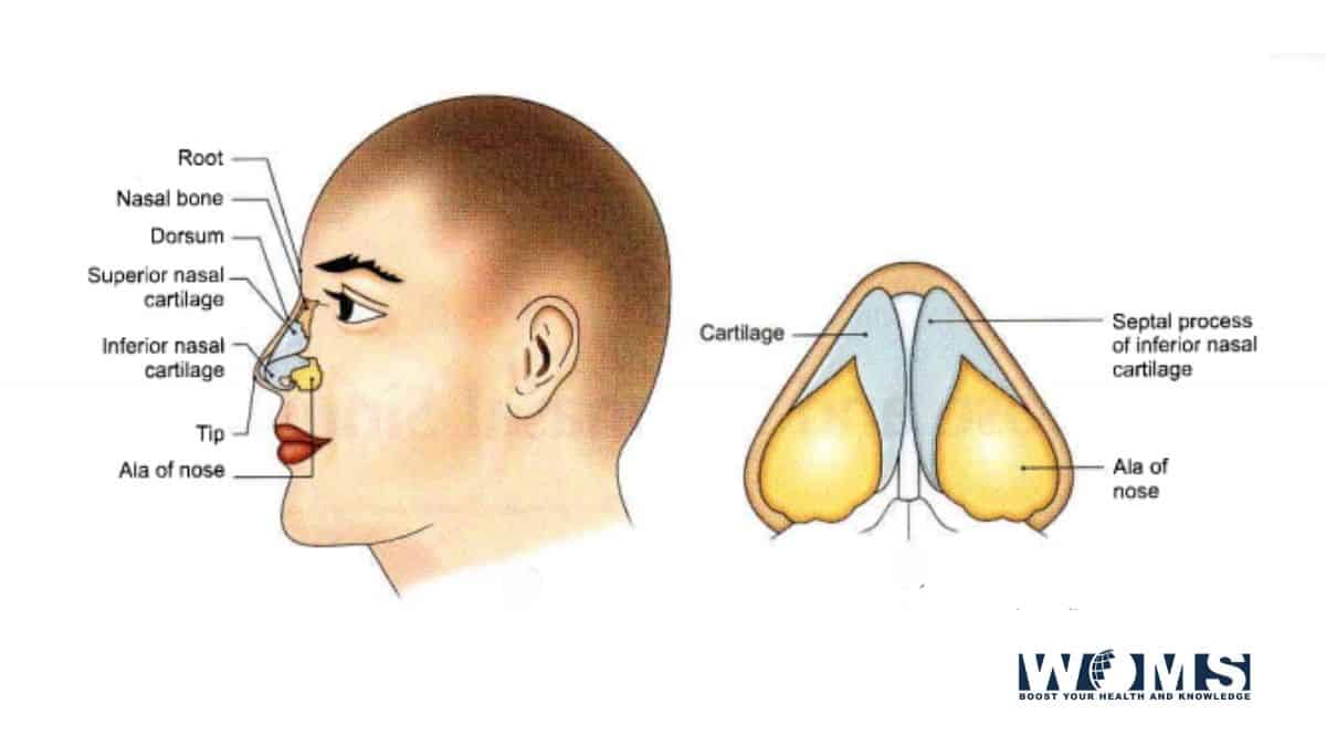
The anatomy of the nose begins with the external nose. The nose is pyramidal in conformation with its root up and the base directed downwards. Various terms are used in its description.
The nasal pyramid consists of an osteocartilaginous framework bound by muscles and skin.
Osteocartilaginous Framework Bony Part
Higher 1/3rd of the external nose is bony while lower 2/3rd are cartilaginous.
The bony part consists of two nasal bones that meet in the midline and rest on the upper part of the nasal process of the frontal bones and are themselves held between the frontal processes of the maxillae.
The anatomy of the nose means the osteocartilaginous framework of the bone.
Cartilaginous Part
It consists of:
- Upper later cartilages: They aggrandize from under the surface of the nasal bones above, to the alar cartilages below. They fuse and with the upper border of the septal cartilage in the midline anteriorly. The lower free edge of the upper lateral cartilage is seen intranasally as limen vestibule, nasal valve, or limen nasi on each side.
- Lower lateral cartilages (alar cartilages). Each alar cartilage is U-shaped. It has a lateral crus that forms the ala and a medical crus which runs in the columella. Lateral crus overlaps the lower edge of the upper lateral cartilage on each side.
- Lesser alar (or sesamoid) cartilages. Two or more in number. They lie above and lateral to alar cartilages. The various cartilages are connected and with the adjoining bones by the perichondrium and periosteum. Most of the free allowance of the nostril is consummate of fibrofatty tissue and not the alar cartilage.
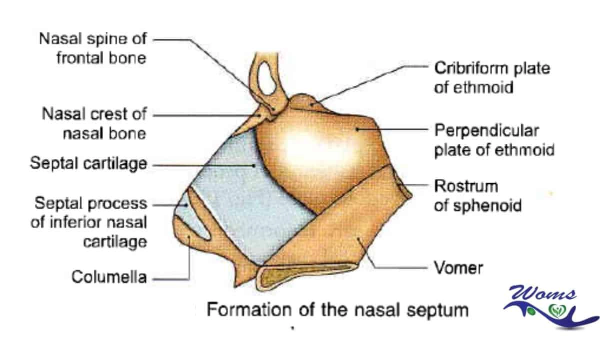
- Septal cartilage. Its anterosuperior border runs from the bottom of the nasal bones to the nasal tip. It abides by the dorsum of the cartilaginous part of the nose. In septal abscess or after excessive removal of septal cartilage as in submucosal resection (SMR) operation, support of nasal dorsum is lost and a supratip depression results.
Nasal Musculature
An osteocartilaginous framework of the nose is covered by muscle which brings about movements of the nasal tip, ala, and the overlying skin. They are the procerus, nasalis (transverse and alar parts), levator labii superioris alaeque nasi, anterior dilator names, and depressor septi.
Nasal Skin
The skin over the nasal bones and upper lateral cartilages is attenuated and advisedly mobile while that covering the alar cartilages is thick and adherent, and contains many sebaceous glands.
It is the hypertrophy of these sebaceous glands which gives rise to a lobulated tumor called rhinophyma.
Anatomy of the Internal Nose
The internal nose is also one of the parts of the anatomy of the nose. It is branched into right and left nasal cavities by the nasal septum.
Each nasal cavity communicates with the exterior through naris or nostrils and with the nasopharynx through the posterior nasal aperture of the choana. Each nasal cavity consists of a skin-lined portion—the vestibule and a mucosa-lined portion, the nasal cavity proper.
Vestibule of Nose
The antecedent and inferior part of the nasal cavity is called the vestibule. It is lined by the skin and bottle up sebaceous glands, hair follicles, and the hair called vibrissae. It’s a higher limit on the lateral wall is prominent by limen nasi (also called nasal valve).
- Nasal valve. It is bounded laterally by the lower border of upper lateral cartilage and fibrofatty tissue and anterior end of inferior turbinate, medially by the cartilaginous nasal septum, and caudally by the floor of pyriform aperture. The angle between the nasal septum and the lower border of the upper lateral cartilage is nearly 30°.
- Nasal valve area. It is the cross-sectional area bounded by the structures forming the valve. It is the least cross-sectional area of the nose and regulates airflow and resistance on inspiration.
Nasal cavity proper
Each nasal cavity has a lateral wall, a medium wall, a roof, and a floor.
Lateral Nasal Wall
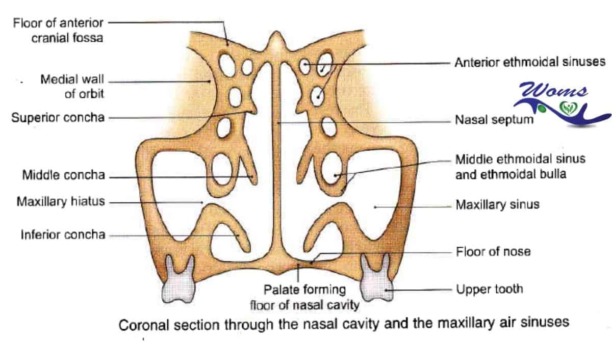
Three and periodically four turbinates or conchae mark the lateral wall of the nose. Nasal conchae or turbinates are scroll-like bony computation covered by mucous membrane. The spaces below the turbinates are called meatuses.
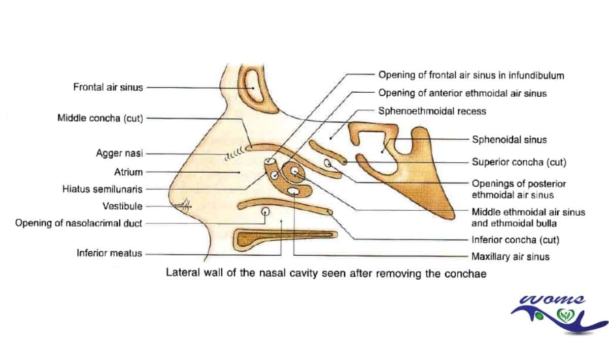
Inferior turbinate: It is a separate bone and below it, into the inferior meatus, opens the nasolacrimal duct guarded at its terminal end by a mucosal valve called Hasner’s valve.
Middle turbinate: It is an ethmoturbinal—a part of the ethmoid bone. It is attached to the lateral wall by a bony lamella called ground or basal lamella. Its attachment is not straight but in an S-shaped manner. In the anterior third, it lies in the sagittal plane and is attached to the lateral edge of the cribriform plate.
In the middle third, it belongs in the frontal plane and is allocate to the lamina papyracea while in its posterior third, it runs horizontally and forms the roof of the middle meatus and is appointed to lamina papyracea and medial wall of the maxillary sinus.
The Ostia of various sinuses draining anterior to basal lamella form the anterior group of paranasal sinuses while those which open posterior and superior to it form the posterior group.
Middle meatus
It shows several important structures that are important in endoscopic surgery of the sinuses.
The uncinate process is a hook-like structure running in the form anterosuperior to the posteroinferior direction. Its posterosuperior border is sharp and runs parallel to the anterior border of bulla ethmoidal; the gap between the two is called hiatus semilunaris (inferior). It is a two-dimensional space of 1-2mm width.
The anteroinferior circumference of the uncinate process is attached to the lateral wall. The posteroinferior borderline of the uncinate process is attached to the inferior turbinate dividing the membranous part of the lower-middle meatus into anterior and posterior fontanelle.
The fontanel area is devoid of bone and consists of membrane only and leads into maxillary sinus when perforated. The upper attachment of the uncinate process shows great variation and may be inserted into the lateral nasal wall, upwards into the base of the skull, or medially into the middle turbinate. This also accounts for variations in the drainage of the frontal sinus.
The space bounded medially by the uncinate process and frontal process of the maxilla and sometimes lacrimal bone, and laterally by the lamina papyracea is called the infundibulum.
The natural ostium of the maxillary sinus is situated in the lower part of the infundibulum. Accessory ostium or Ostia of the maxillary sinus are constantly seen in the anterior or posterior fontanel.
Bulla ethmoidal
It is an ethmoidal cell situated behind the uncinate process. The anterior surface of the bulla composes the posterior boundary of hiatus semilunaris.
Depending on pneumatization, bulla may be a pneumatized cell or a solid bony prominence. It may extend superiorly to the skull base and posteriorly to fuse with ground lamella. When there is a space above or behind the bulla, it is called suprabulla or retrobulbar recesses, respectively.
The suprabullar and retrobulbar adjourn together to form the lateral sinus (sinus lateral of Grunwald). The lateral sinus is thus encompassed superiorly by the skull base, laterally by lamina papyracea, medially by middle turbinate.
The cleft-like communication between the bulla and skull base and opening into the middle meatus is also called hiatus semilunaris superior in contrast to hiatus semilunaris inferior referred to before.
Atrium of the middle meatus:
It is a shallow depression lying in front of the middle turbinate and above the nasal vestibule. you must learn detail about the meatus in the anatomy of the nose.
Agger nasal:
It is an elevation just anterior to the attachment of the middle turbinate. When pneumatized it contains air cells, the agger nasi cells, which communicate with the frontal recess.
A broadened agger nasi cell may impinge on the frontal recess area, constricting it and causing mechanical blocking to frontal sinus drainage.
Pneumatization of middle turbinate leads to an enlarged ballooned out middle turbinate called concha bullosa. It cesspool into frontal recess directly or through agger nasi cells.
Haller cell is air cells situated in the roof of the maxillary sinus. They are pneumatized from anterior or posterior ethmoid cells. Enlargement of Haller cells encroaches on ethmoid infundibulum, impending draining of maxillary sinus.
Superior turbinate: It is also an ethmoturbinal and is situated posterior and superior to the middle turbinate. It may also pneumatized by one or more cells. It forms an important landmark to identify the ostium of the sphenoid sinus which lies medial to it.
Superior meatus: It is a space below the superior turbinate. Posterior ethmoid cells open into it. The number of posterior ethmoid cells varies from 1 to 5.
Onodi cell is a posterior ethmoidal cell that may grow posteriorly by the side of the sphenoid sinus or superior to it for as much distance as 1.5 cm from the anterior surface of the sphenoid. Onodi cell is surgically important as the optical nerve may be related to its lateral wall.
Sphenoethmoidal recess: It is situated above the superior turbinate. Sphenoid sinus opens into it.
Supreme turbinate: It is sometimes nonce above the superior turbinate and has a narrow meatus beneath it. The ostium of the sphenoid sinus is situated in the sphennoethmoidal recess medial to the superior or supreme turbinate. It can be occupying endoscopically about 1cm above the upper margin of the posterior choana close to the posterior border of the septum.
Medial Wall
The nasal septum forms the medial wall.
Roof
The anterior acclivous part of the roof is formed by nasal bones, the posterior sloping part is formed by the body of the sphenoid bone and the middle horizontal part is formed by the cribriform plate of the ethmoid through which the olfactory nerves enter the nasal cavity.
Floor
It is arranged by the palatine process of the maxilla in its anterior three-fourths and horizontal part of the palatine bone in its posterior one-fourth.
Kiesselbach plexus
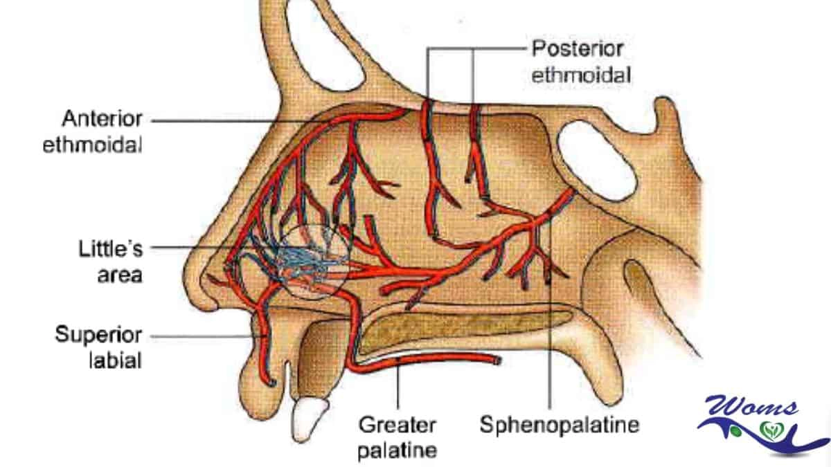
Kiesselbach plexus, also known as the little’s area, is a vascular anastomosis of the four arteries. The kiesselbach plexus is referred to as kiesselbach’s triangle or kiesselbach’s area. The Littles area is the stock site for epistaxis (nasal bleeding).
The four arteries that the kiesselbach plexus includes are:
- Anterior ethmoidal artery-Branch of the ophthalmic artery)
- Sphenopalatine artery-terminal branch of the maxillary artery)
- Greater palatine artery-from the maxillary artery)
- Septal branch of the superior labial artery-from the facial artery)
The clinical importance of the kiesselbach plexus is epistaxis. Ninety percent of nasal bleeding occurs in this area as it is exposed to the drying area, nail trauma. Nasal bleeding is common for both in case of children and adults.
The lining membrane of the internal nose
- VESTIBULE. It is lined by skin repressing hair, hair follicles, and sebaceous glands.
- OLFACTORY REGION. Upper one-third of the lateral wall (up to superior concha), corresponding part of the nasal septum, and the roof of the nasal cavity from the olfactory region. In this region, the mucous membrane is paler in color.
- RESPIRATORY REGION. Lower two-thirds of the nasal cavity from the respiratory region. Here in the respiratory region mucous membrane conduct variable thickness being thickest over nasal conchae conspicuously at their ends, quite thick over the nasal septum but very thin in the meatuses and floor of the nose. It is highly vascular and also contains erectile tissue. Its surface is lined by pseudostratified ciliated columnar epithelium which contains plenty of goblet cells. In the submucosa layer of the mucous membrane are situated serous, mucous, both serous and mucous secreting glands, the duct of which open on the surface of the mucosa. The lining membrane of the nose is also included as part of the anatomy of the nose.
Nasal conchae
Nasal conchae also called turbinate, or Turbinal, any of several thin, scroll-shaped bony elements forming the upper chambers of the nasal cavities.
Nasal concha increases the surface area of these cavities, thus providing for rapid warming and humidification of air as it passes to the lungs.
Nerve supply
When you start reading the anatomy of the nose, the Nerve supply is one of the important contributions to the anatomy of the nose. Here begins the nerve supply of the nose.
-
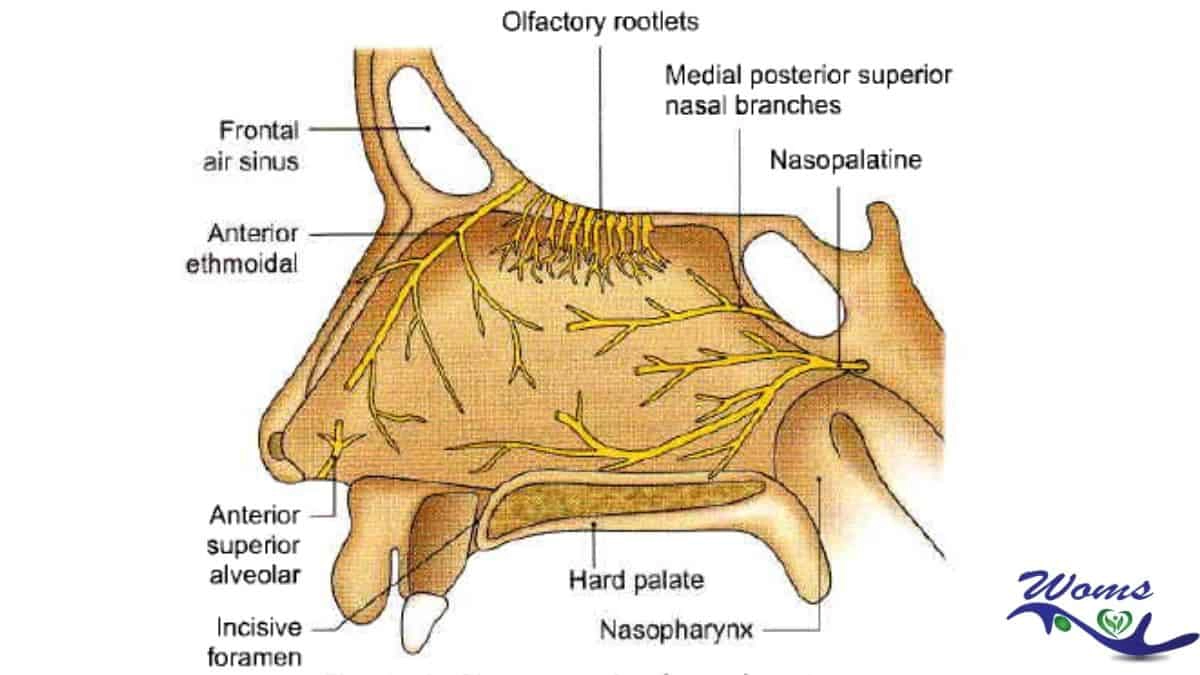
OLFACTORY NERVES:
They cart sense of smell and supply the olfactory region of the nose. They are the central filaments of the olfactory cells and are arranged into 12-20 nerves which pass through the cribriform plate and end in the olfactory bulb.
These nerves can carry sheaths of dura, arachnoid, and pia with them into the nose. Injury to these nerves can open CSF space leading to CSF rhinorrhoea or meningitis.
2. NERVES OF COMMON SENSATION:
They are:
- The anterior ethmoidal nerve.
- Branches of the sphenopalatine ganglion.
- Branches of the infraorbital nerve. They source the vestibule of the nose both on its medial and lateral side.
Most of the posterior two-thirds of the nasal cavity (both septum and lateral wall) are supplied by branches of sphenopalatine ganglion which can be blocked by placing a pledget of cotton soaked in anesthetic solution near the sphenopalatine foramen situated at the posterior extremity of the middle turbinate.
The anterior ethmoidal nerve which supplies the anterior and superior part of the nasal cavity (lateral wall and septum) can be blocked by placing the pledget high up on the inside of nasal bones where the nerve enters.
3.AUTONOMIC NERVES.
Parasympathetic nerve fibers supply the nasal glands and control nasal secretion. They come from the greater superficial petrosal nerve, travel in the nerve of the pterygoid canal (vidian nerve), and reach the sphenopalatine ganglion where they relay before reaching the nasal cavity. They also surplus the blood vessels of the nose and cause vasodilation.
Sympathetic nerve fibers come from the upper two thoracic segments of the spinal cord, pass through the superior cervical ganglion, travel in the deep petrosal nerve, and add the parasympathetic fibers of the greater petrosal nerve to anatomy the nerve of the pterygoid canal (vidian nerve).
They ambit the nasal cavity without relay in the sphenopalatine ganglion. Their simulation causes vasoconstriction. Excessive rhinorrhoea in cases of vasomotor and allergic rhinitis can be controlled by a section of the vidian nerve. The anatomy of the nose also includes the nerve supply of the nose.
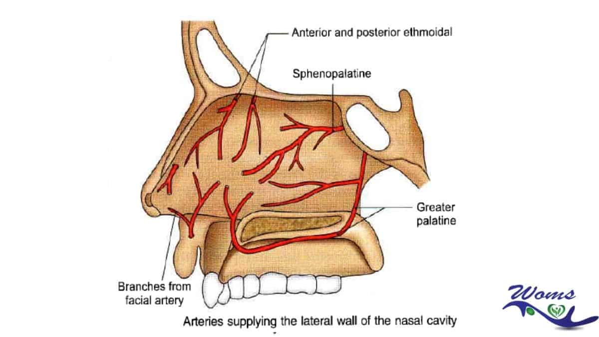
Blood supply
The blood supply is one of the important parts of the anatomy of the nose. Both the internal and external carotid system supply the nose.
Lymphatic Drainage
Lymphatics from the external nose and anterior part of the nasal cavity drain into submandibular lymph nodes while those from the rest of the nasal cavity drain into upper jugular nodes either directly or through the retropharyngeal nodes.
Lymphatics of the upper part of the nasal cavity communicate with subarachnoid space along the olfactory nerves.
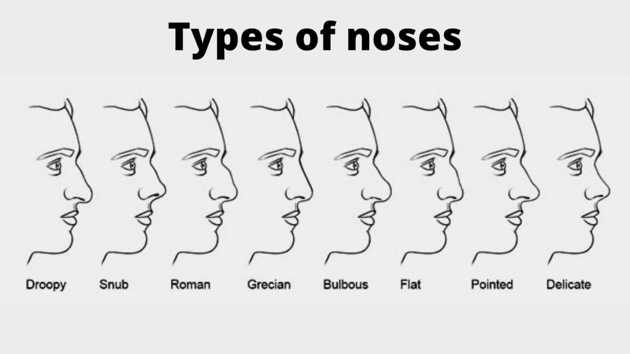
Types of noses
Here are eight types of noses:
- Types of Noses: the Nubian nose
- The greek nose
- The hook nose
- The arched nose
- The button nose
- The straight nose
- The concave nose
- Types of Noses: the crooked nose.
Also Read about: Anatomy of the heart
Conclusion
The human nose is the organ that helps in breathing as well as smell. Concerning nose anatomy, the inner portion of the nose lies above the roof of the mouth.
The anatomy of the nose includes the external meatus, external nostrils, septum, nasal passage, and sinuses. The nasal bones form the important part of the nose.
The external meatus is a triangular-shaped projected structure. It lies in the center of the face. There are two outer chambers known as external nostrils. They are divided by the septum.
Another part of nose anatomy is the septum. Anatomically, it is made of bone as well as cartilage. It remains covered by the mucous membrane.
The passage of the nose that filters the air is a nasal passage. It consists of tiny hairs called cilia and is lined with mucous membranes. Sinuses are also lined with mucous membranes. They are air-filled cavities present in four pairs.
