Kussmaul’s Sign

Kussmaul’s sign indicates an increase in the jugular venous pressure (pressure during filling of the jugular vein) on inspiration. This abnormal sign is because of failure to fall in venous pressure of the jugular vein with the inspiration process.
History of Kussmaul’s sign
In 1873, Kussmaul explained this phenomenon, discovered by Greisinger in 1854, in which he described jugular veins becoming distended with each inspiration. This sign is opposite to the normal physiological process that is jugular venous pressure (JVP) falls with inspiration. He also has credits to explain Kussmaul’s breathing process.
Pathophysiology of positive Kussmaul’s sign
Generally, the jugular venous pressure falls with inspiration as the pressure reduces in the expanding thoracic cavity. The negative intrathoracic pressure sucks in the blood and reduces the venous pressure. In contrast, Kussmaul’s sign appears because of the failure to fall in this venous pressure. This sign more frequently occurs because of various malfunctioning and constrictive diseases.
The pathophysiological mechanisms that result in Kussmaul’s sign are based on such conditions which cause:
- Right ventricular dysfunction
- Impaired filling of the right ventricle
- Increased right atrial pressure
There are three fundamental disorders that act as a barrier to the effective filling of the right ventricle. As a result, this causes a paradoxical increase in jugular venous pressure with inspiration. These significant factors are as follows to describe the pathophysiological phenomenon of Kussmaul’s sign.
- Decreased elasticity or inelasticity of myocardium due to fibrosis and infections (restrictive cardiomyopathy)
- Constrictive pericardial diseases (constrictive pericarditis)
- Disturbed right ventricular function due to right myocardial infarction
How to determine the manifestation of Kussmaul’s sign?
There are two methods to assess the behavior of Kussmaul’s sign.
Invasive method
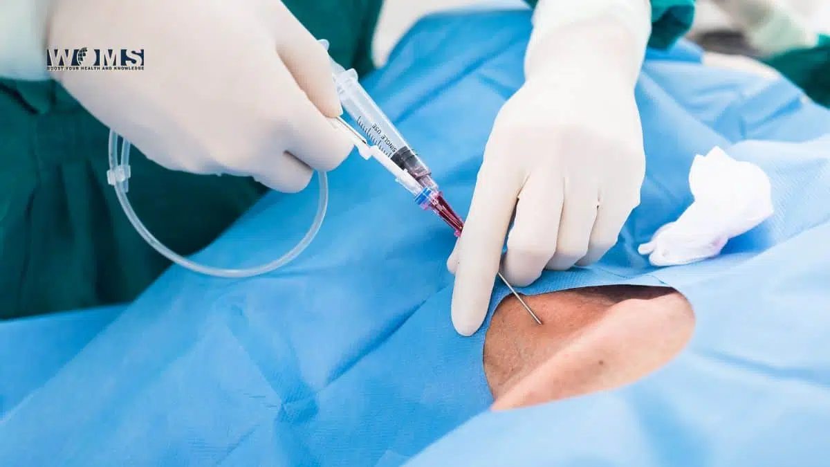
This method is the measurement of central venous pressure. Central venous pressure approximates the right atrial pressure. If it raises during inspiration, it indicates an impaired venous return to the right heart. This finding is indicative of positive Kussmaul’s sign.
Non-invasive method:
This is the most common method for the evaluation of right ventricular filling pressure. It only uses a simple flashlight and a ruler. The increased jugular venous pressure in comparison to pre-inspiration represents Kussmaul’s sign.
The differential diagnosis for positive Kussmaul’s sign:
This sign is indicative of various diseases related to the dysfunction of the right heart. In addition, Kussmaul’s sign also highlights various pulmonary disorders associated with heart disturbances. Some of these are significant diseases with positive Kussmaul’s sign.
Constrictive pericarditis
In this condition, Kussmaul’s sign is positive because of the less compliant pericardium. The pericardium restricts the diastolic filling of the right ventricle, causing increased atrial pressure. These conditions act synergistically and result in a paradoxical rise in jugular venous pressure, known as Kussmaul’s sign.
Restrictive cardiomyopathy
Restrictive cardiomyopathy also induces the impaired filling of the right ventricle. As a result, it will increase the pressure of the right atrium. In this way, there will be a rise in jugular venous pressure indicative of Kussmaul’s sign.
Right heart failure
Right heart failure represents the dysfunction of the right-sided heart. As a result, it will disturb the filling of the right ventricle, leading towards increased JVP.
Right-sided heart tumor
Any tumor of the right-sided heart will lead towards the dysfunction of the right heart. This tumor will obstruct the proper flow of the venous blood. In addition, it will raise the JVP indicating Kussmaul’s sign.
Fibrosis or stenosis of the tricuspid valve
Fibrosis or stenosis of the tricuspid valve obstructs the flow from the right atrium to the right ventricle. It will increase atrial pressure and increase JVP.
Right-sided predominant heart infarction
Right-sided heart infarction disturbs the normal function of the right heart. As a result, it leads towards increased JVP.
Massive pulmonary embolism
Pulmonary embolism is also an etiologic factor for Kussmaul’s sign. The embolus will obstruct the flow from the right ventricle to the lungs leading towards increased atrial pressure as well as JVP.
Jugular venous pressure(JVP) waveform
Jugular venous pressure is the measurement of central venous pressure. This pressure represents the venous pressure via direct visualization of the internal jugular vein. This measurement helps to differentiate between different heart and lung diseases. Let’s have a view of normal and abnormal jugular venous pressure in multiple diseases.
The normal waveform for the jugular venous pressure represent as follows:
a – atrial contraction
x – atrial diastole
c – the ballooning of tricuspid valve with ventricular contraction
x’ – down movement of tricuspid valve with ventricular contraction
v – passive filling of the right atrium
y – right atrium clearing with the opening of the tricuspid valve
(a, c, v – waves) (x, y – descents)
Diseases associated with Kussmaul’s sign (increased JVP)
- Constrictive pericarditis shows prominent and deepy-descent
- Right heart failure shows a largea-wave which indicates increased atrial contraction pressure.
- Pulmonary hypertension also exhibits large a-wave
- Tricuspid stenosis exhibits slow y-descent and large a-wave
Correction of positive Kussmaul’s sign
Kussmaul’s sign is an indication of any underlying heart or lung disease, not a disease itself. Therefore, this condition requires the management of the underlying diseases. For that, you have to undergo various clinical as well as diagnostic examinations. Kussmaul’s sign is a symptom of various diseases. This symptom disappears with the management of the underlying disease.
FAQs
How to measure JVP?
● Position the patient in a semi-reclined position at a 45° angle.
● Turn the patient’s head slightly towards the left.
● Inspect the internal jugular vein between the medial end of the clavicle and ear lobe.
● Measure the JVP by evaluating the vertical distance between the sternal angle and the pulsation point of the internal jugular vein.
What is pseudo-Kussmaul’s sign?
This is an observation of severe respiratory insufficiency. This condition mimics the finding of Kussmaul’s sign. But, the main cause is the mechanical obstruction of the external jugular vein, not any heart or lung disease.


