Dengue Fever: what all you must know
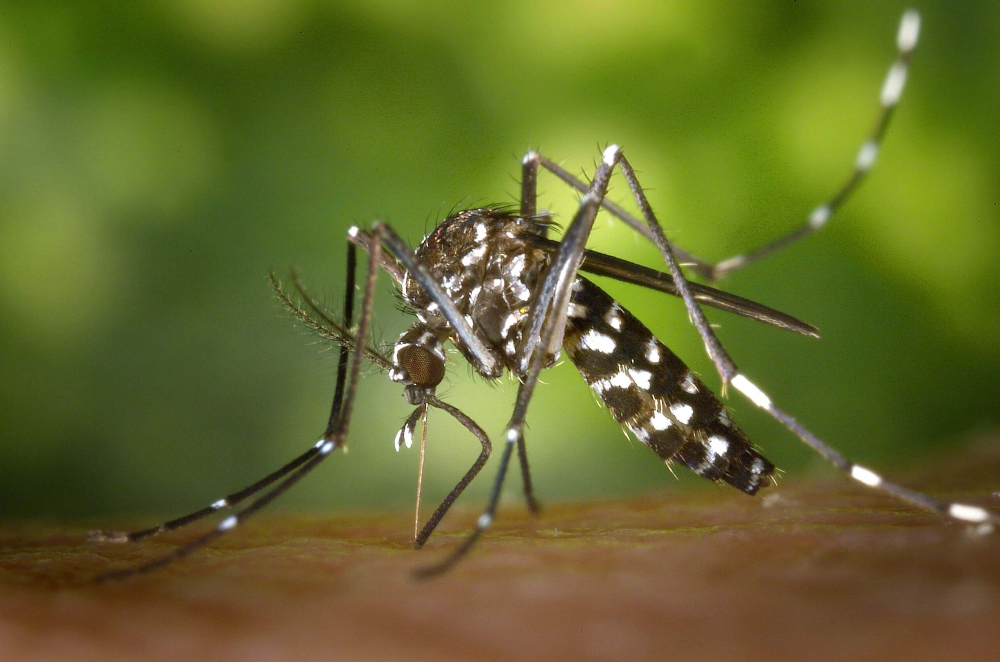
Dengue fever is a febrile illness caused by a flavivirus transmitted by mosquitoes. It is endemic in Asia, the Pacific, Africa, and the Americas. Approximately 400 million infections and 100 million clinically apparent infections occur annually, and dengue fever is the most rapidly spreading mosquito-borne viral illness. The principal vector is the mosquito Aedes aegypti, which breed in standing water; a collection of water in containers, water-based air coolers, and tire dumps are good environments for the vector in large cities.
Aedes albopictus is a vector in some south-east Asian countries. There are four serotypes of the dengue virus, all producing a similar clinical syndrome; type-specific immunity is life-long but immunity against the other serotypes lasts only a few months. Dengue hemorrhagic fever (DHF) and dengue shock syndrome (DSS) occur in individuals who are immune to one dengue virus serotype and are then infected with another. Prior immunity results in increased uptake of the virus by cells expressing the antibody Fc receptor and increased T-cell activation with resultant cytokines release, causing a capillary leak and disseminated intravascular coagulation.
Previously, Dengue was seen in small children and DHF/DSS in children 2-15 years old, but these conditions are now being seen in children less than 2 years old, and most frequently in those 16-45 years of age or older, in whom severe organ dysfunction is more common. Other epidemiological changes include the spread of dengue into rural communities and greater case fatality in women.
Clinical features
Many cases of dengue infections are symptomatic in children, the clinical disease presents with undifferentiated fever termed dengue-like illness. When dengue infection occurs with characteristics symptoms or signs it is termed ‘dengue’ or ‘dengue fever’. A rash frequently follows the initial febrile phase as the fever settles. Laboratory features include leucopenia, neutropenia, thrombocytopenia, and elevated alanine aminotransferase (ALT) or aspartate aminotransferase (AST).
Many symptomatic infections run an uncomplicated course but complications or a protracted convalescence may ensure. Warning signs justify intense medical management and monitoring for progression to severe dengue, Atypical clinical features of dengue are increasingly common, especially in infants or older patients. These, along with DHF or DHSS, are recognized as features of severe dengue fever in the 2015 case definition.
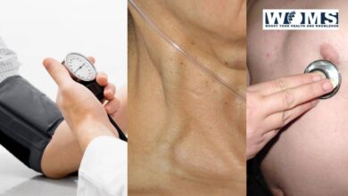
The period of 3-7 days after onset of fever is termed the ‘critical’ phase, during which signs of DHF or DSS may develop. In mild forms, petechiae occur in the arm when a blood pressure cuff is inflated to a point between systolic and diastolic blood pressure and left for 5 minutes (the positive tourniquet test)- a non-specific test of capillary leak increases, DSS develops, with a raised hematocrit, tachycardia, and hypotension, pleural effusions, and ascites.
This may progress to metabolic acidosis and multi-organ failure, including acute respiratory distress syndrome (ARDS). Minor (petechiae, ecchymoses, features of DHF, may occur, cerebrovascular bleeding may be a complication of severe dengue fever.
Incubation period
2-7 days
Prodrome
2 days of malaise and headache
Acute onset
- Fever
- Backache
- Arthralgias, headache, generalized pains (‘break-bone fever’)
- Pain on eye movement
- Lacrimation
- Scleral injection
- Anorexia
- Nausea and vomiting
- Pharyngitis, upper respiratory tracts symptoms
- Relative bradycardia
- Prostration
- Depression
- Hyperaesthesia
- Dysgeusia
- Lymphadenopathy
Fever
Continous or ‘saddle-black’, with a break on 4th or 5th day and then recrudescence; usually last 7-8 days
Rash
Initial mantling faint macular rash in first 1-2 days. Maculopapular, scarlet morbilliform blanching rash from days 3-5 on the trunk, spreading centrifugally and sparing palms and soles; onset often with fever dereference. May desquamate on dauntlessness or give rise to petechiae on extensor surfaces.
Convalescence
Slow and may be associated with prolonged fatigue syndrome, arthralgia or depression
Complications
- Dengue hemorrhagic fever and disseminated intravascular coagulation
- Dengue shock syndrome
- Severe organ involvement
- Vertical communication if infection within 5 weeks of delivery
WHO case definition of dengue, 2015
Probable dengue fever
- Exposure in an endemic area
- Fever
- Two of
- Nausea/vomiting
- Rash
- Aches/pains
- Positive tourniquet test
- Leucopenia
- Any warning sign
Laboratory confirmation important
Needs regular medical ascertainment and instruction in the warning signs
If there are no warning signs, the need for hospitalization is influenced by age, comorbidities, pregnancy and social factors
Dengue with warning signs
- Probable dengue plus one of:
- Abdominal pain or tenderness
- Persistent vomiting
- Sign of fluid accumulation, e.g. pleural effusion or ascites
- Mucosal bleed
- Hepatomegaly>2 cm
- The rapid increase in hematocrit with a fall in platelet count
Need medical intervention, e.g. intravenous fluid
Severe dengue
- Severe plasma leakage leading to:
Shock(dengue shock syndrome)
Fluid accumulation with respiratory distress
- Severe hemorrhagic manifestation, e.g. gastrointestinal hemorrhage
- Severe organ involvement (atypical features):
Liver AST or ALT ≥1000 U/L
CNS: impaired consciousness, meningoencephalitis, seizures cardiomyopathy, conductions defects, arrhythmias
Other organs, e.g. acute kidney injury, pancreatitis, acute lung injury, disseminated intravascular coagulopathy, rhabdomyolysis
Needs exigency medical treatment and specialist care with intensive care input.
Diagnosis
In endemic areas, mild dengue must be distinguished from other viral infections. The diagnosis can be confirmed by seroconversions of IgM or a fourfold rise in IgG antibody titers. Serological tests may detect cross-reacting antibodies from infection or vaccination against flaviviruses, including yellow fever virus, Japanese encephalitis virus, and West Nile virus. Isolation of dengue virus or detection of dengue virus by RNA by PCR in blood or CSF is available in specialist laboratories. Commercial enzyme-linked immunosorbent assay ELISA kits to detect the nS1 viral antigen, although less sensitive than PCR, are available in many endemic areas.
Management and Prevention
Treatment is supportive, emphasizing fluid replacement and appropriate management of shock and organ dysfunction, which is a major determinant of morbidity and mortality. With intensive care abutment, mortality rates are 1% or less. Aspirin should be avoided due to bleeding risk. Glucocorticoids have not been shown to help. No existing antivirals are effective.
Breeding places of Aedes mosquitoes should be abolished and the adults destroyed by insecticides. A recently licensed vaccine is available.
