Vagina Pics with its anatomy
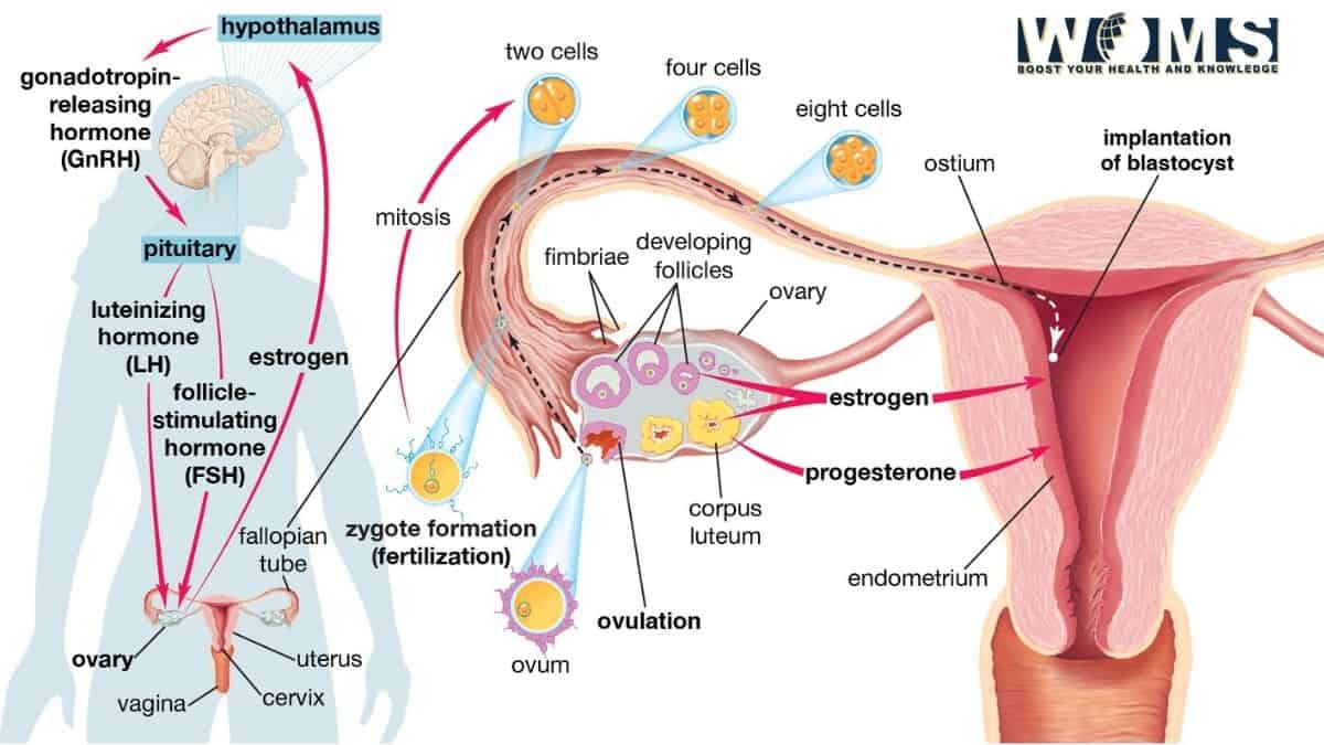
The vagina is an important reproductive organ in females. The word ‘vagina’ means a sheath. It is the fibro-muscular canal, which forms the female Copulatory organ. In this post, you are going to learn about the anatomy of the vagina, and you can also visualize different vagina pics. There are vagina pics to assist you in understanding the topic better.
The female reproductive organ vagina prolongates from the vulva to the uterus. It is present behind the bladder and the urethra and in front of the rectum and anal canal.
Vagina anatomy
Let’s begin the anatomy of the vagina with its shape and size. The anterior wall of the vagina is about eight centimeters long. And the posterior wall of the vagina is about ten centimeters long.
Relations of the anterior wall
- The upper half of the anterior wall is related to the base of the urinary bladder.
- The lower half of the urethra
Relations of the posterior wall
- The Upper one-fourth of the posterior wall of the vagina is apart from the rectum by the rectouterine pouch.
- The middle two-fourths are abstracted from the rectum by loose connective tissue.
- The lower one-fourth of the posterior wall isn’t in connection to the anal canal. It is separated by the perineal body and the muscle attached to it.
Anatomical vagina pictures
While learning anatomy pictures play a vital role. Similarly to understand the vagina, the vagina pics are vital. Here are some of the vagina pics which will help you a better understanding.
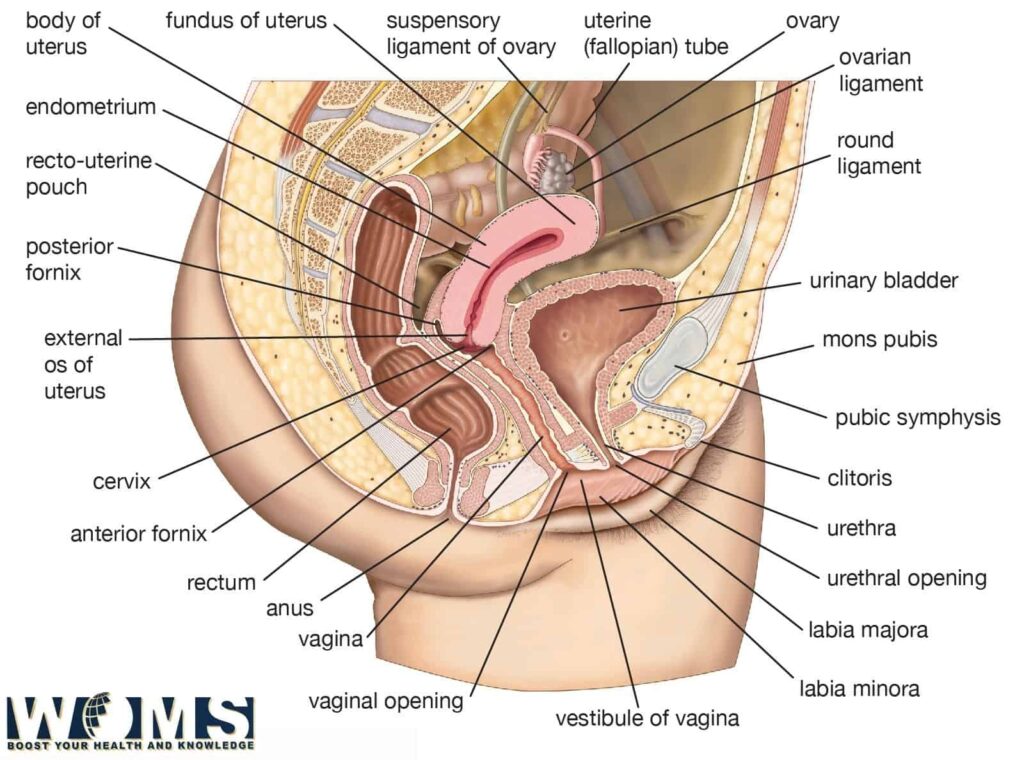
The diameter of the vagina slowly increases from below upwards. The upper end of the vault is roughly five centimeters twice the size of the lower end which is 2.5 centimeters. However, it is absolutely extensive and allows passage of the head of the fetus during normal delivery.
Anatomy of the vagina in virgin
In virgin women, the lower end of the vagina is incompletely closed by a fine annular fold of mucous membrane called the hymen.
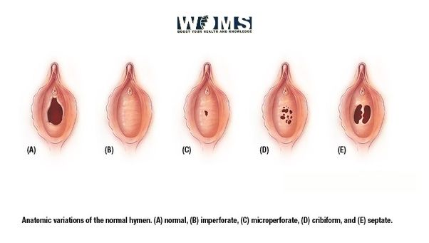
In married women, the hymen is characterized by circular acclivity around the vaginal orifice, the caruncular hymenales.
Arterial supply of vagina
The vagina is also a vascular organ, and the following arteries supply it:
- The principal artery supplying is the vaginal branch of the internal iliac artery.
- The cervicovaginal branch of the uterine artery supplies the upper part. The middle rectal and internal pudendal arteries supply the lower part.
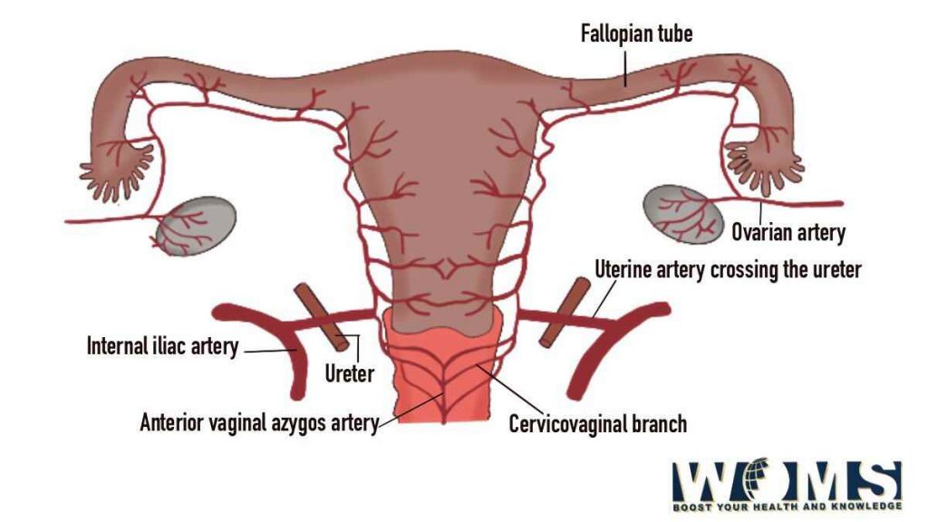
Clinical anatomy of the vagina
Inspection
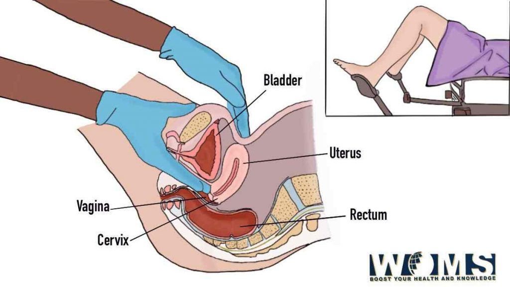
The vagina is first inspected at the introitus by separating the labia minora with the left hand. Next, the speculum examination helps to inspect the cervix and vaginal vault and to take the vaginal swab.
Palpation
A pr vaginal (PV) digital examination helps to do palpation of the pelvic organs. With the examining fingers one can feel:
- Anteriorly: the urethra, bladder, and pubic symphysis.
- Posteriorly: the rectum and pouch of Douglas.
- Laterally: the ovary, tube, lateral pelvic wall, thickened ligaments, and ureters.
- Superiorly: the cervix
Bimanual (abdominovaginal) examination helps in the assessment of the size and position of the uterus, enlargements of the ovaries and tubes, and other pelvic masses.
Vagina conditions with vagina pics
Vaginal lacerations: Traumatic lacerations of the vagina are common, and may be due to forcible coitus (rape), during childbirth, or accidents. This may give rise to profuse bleeding.
Vaginitis: it is common before puberty and after menopause because of the thin delicate epithelium. In adults, the resistant squamous epithelium prevents infections.
The trichomonas vaginalis, and gonococcal mainly cause Vaginitis. It causes leucorrhea or white discharge.
Vaginal prolapsed: prolapse of the anterior wall of the vagina drags the bladder (cystocele) and urethra (Urethocoele), and the posterior wall drags the rectum (rectocele).
The weakness of supports of the uterus can give rise to different degrees of prolapse. Similarly, trauma to the anterior and posterior walls of the vagina can cause vesicovaginal, urethrovaginal, and rectovaginal fistulae.
Vaginal cancer: Primary new growths of the vagina, like infections, are uncommon. However, the secondary involvement of the vagina by the cancer cervix is common.
Vaginal test
- Pelvic examination: using a speculum, a gynecologist can examine the vulva, vagina, and cervix. This examination helps to test the strength of the pelvic muscle.
- Pap smear cytology: pap tests are cytologic preparations of exfoliated cells from the cervical transformation zone that are stained with the Papanicolaou method.
- Colposcopic examination and VIA (Visual inspections with acetic acid)
- Vaginal biopsy: Histologic diagnosis and removal of precancerous lesions.
Vaginal Treatment
Antimicrobials drugs: These are the substances produced by microorganisms, which suppress the growth or kill of other microorganisms at very low concentrations. Antifungal medications like Miconazole, Clotrimazole, and nystatin can treat yeast infections. For bacterial infections, you can use antibiotics. Antiviral medicines like acyclovir, gancyclovir, famciclovir, and more can treat the herpes virus infections
Wart treatment: Freezing, chemicals, burning with laser, or cautery are some methods used for the treatment of the wart. Do read about the home remedies for warts.
Exercises: Kegel exercises help to strengthen the pelvic muscle which helps to improve or prevent vaginal prolapse. It may also prevent urinary incontinence.
Estrogen: In the treatment of different vaginal conditions, estrogen can be used. Both the internal and external organs of females respond to estrogen.
Surgery: Surgery is required for the differentiation of the tumor. It is also used for the treatment of vaginal prolapse.
Takeaway:
The vagina is an important female reproductive organ. It is a fibromuscular canal. The vagina suffers from different conditions like vaginitis and vagina prolapse. There are vagina pics showing the different vagina conditions. There are various treatments for the vagina conditions like using antimicrobial drugs or doing surgery in extreme cases.
