Anatomy of the lungs
Anatomy of the Lungs: Introduction and some features
Lungs are the vital organ of the Respiratory system. There are two lungs present in the human body. Lungs help us to exchange gases from the environment. Lungs are the organ that helps in inhaling oxygen and expiring carbon dioxide. To know about the lungs, we must understand the anatomy of the lungs. Let’s also know about bronchopulmonary segments, lung parenchyma, and lung hilum.
The lungs are occupying significant portions of the thoracic cavity, leavers little space for the heart, which excavates more of the left lung. The two lungs hold the heart tight between them, providing it with the protection it rightly deserves. There are ten bronchopulmonary segments in each lung.
The lungs are a pair of respiratory organs situated in the thoracic cavity. Each lung invaginates the corresponding pleural cavity. The mediastinum separates the right and left lungs.
The lungs are spongy in texture. In the young, the lungs are brown or grey. Gradually, they become mottled black because of the deposition of inhaled carbon particles. The right lung weighs about 700g; it is about 50 to 100 g heavier than the left lung.
Hilum of the left lungs shows the single bronchus situated posteriorly, with bronchial vessels and posterior pulmonary plexus. The pulmonary artery lies above the bronchus. Anterior to the bronchus is the upper pulmonary vein, while the lower vein lies below the bronchus.
The mediastinal surface of the left lung has the impression of the left ventricle, ascending aorta. Behind the root of the left lung are impressions of descending thoracic aorta while the esophagus leaves an impression in the lower part only. External features of the lungs are one of the main intrinsic elements of the anatomy of the lungs.
Features of the lungs
Both of the anatomy of the lungs are conical in shape and has the following features:
- An apex at the upper end.
- A base is resting on the diaphragm.
- Three borders of the lungs are anterior, posterior, and inferior borders.
- Two surfaces of the lungs are costal and medial. The medial surface divides into vertebral and mediastinal parts.
The apex is blunt and lies above the level of the anterior end of the first rib. It reaches nearly 2.5 cm above the medial one-third of the clavicle, just medial to the supraclavicular fossa. It is covered by the cervical pleura and by the suprapleural membrane, and grooved by the subclavian artery on the medial side and anteriorly.
The base is semilunar and concave. It rests on the diaphragm which separates the right lung from the right lobe of the liver, and the left lung from the left lobe of the liver, the fundus of the stomach, and the spleen.
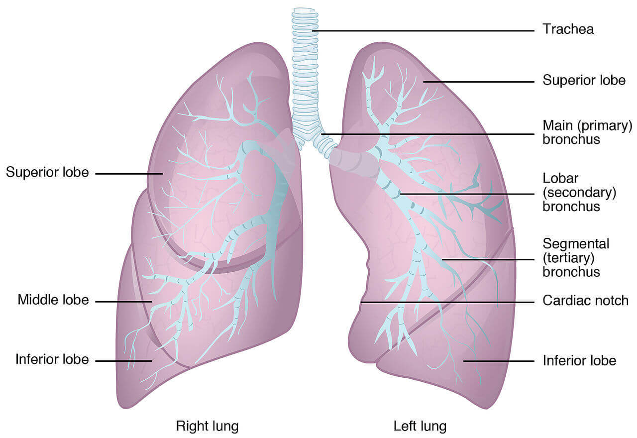
The anterior border is very thin. It is shorter than the posterior border. On the right side, it is vertical and corresponds to the anterior or costomediastinal line of pleural reflection. The anterior border of the left lung shows a wide cardiac notch below the level of the fourth costal cartilage. The heart and pericardium don’t cover by the lung in the region of the notch.
The posterior border is thick and ill-defined. It corresponds to the medial margins of the heads of the ribs. It extends from the level of the seventh cervical spine to the tenth thoracic spine.
The inferior border abstracted the base from the costal and medial surfaces.
The costal surface is large and convex. It is in contact with the costal pleura and the overlying thoracic wall.
The medial surface divides into a posterior or vertebral part and an anterior or mediastinal part. The vertebral part is related to the structures like vertebral bodies, intervertebral disc, the posterior intercostal vessels, and the splanchnic nerves. The mediastinal part is associated with the mediastinal septum and shows a cardiac impression, the hilum, and several other impressions that differ on the two sides.
Fissures and lobes within the anatomy of the lungs
The right lung is divided into three lobes (upper, middle, and lower) by two fissures, oblique and horizontal. The left lung is branched into two lobes by the oblique fissure.
The oblique fissure cuts into the whole thickness of the lung, except at the hilum. It passes obliquely downwards and forwards, crossing the posterior border about 6cm below the apex and the inferior border about 5cm from the medial plane. Due to the oblique plane of the fissure, the lower lobe is more posterior and the upper and middle lobe more anterior.
In the right lung, the recumbent fissure passes from the anterior border up to the oblique fissure and separates a wedges-shaped middle lobe from the upper lobe. The fissure runs horizontally at the level of the fourth costal cartilage and meets the oblique fissures in the midaxillary line.
The tongue-shaped projection of the left lung below the cardiac notch is called the lingula. It assimilates to the middle lobe of the right lung.
The lungs expand maximally in the inferior direction because movements of the thoracic wall and diaphragm are maximal towards the base of the lung. The being of the oblique fissure of each lung allows a more uniform expansion of the whole lung.
Bronchopulmonary segments:
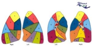
If the part of the anatomy of the lungs that are supplied by specific segment bronchus and arteries then it is called Bronchopulmonary segments. It can be also defined as, If the lung is divided into segments based on tertiary bronchus then it is called Bronchopulmonary segments. The segments are separated from one another by connective tissue. So, thus they are also called as an anatomical or functional unit. There are ten bronchopulmonary segments in the right lung while there are eight to nine bronchopulmonary segments in the left lung.
Lung Parenchyma:
Lung parenchyma is also known as the pulmonary parenchyma which is the portion of the lungs involved in the gaseous transfer. The lung parenchyma consists of the alveoli, alveolar ducts, and the respiratory bronchioles. Each alveolus in the lung parenchyma opens directly into alveolar ducts. Sometimes in a limited number, lung parenchyma can open into a respiratory bronchiole too.
Lung Hilum:
Lung hilum is present on the medial aspect of the lungs. It is through the lung hilum the other structure like veins, artery, etc enter and leave the lungs. It is also the point for the attachment of lung root.
The root of the lungs
The root of the anatomy of the lungs is a short, broad pedicle that connects the medial surfaces of the lung to the mediastinum. It is formed by structures that either enter or come out of the lung at the hilum (Latin depression). The roots of the lungs lie opposite the bodies of the fifth, sixth, and seventh thoracic vertebrae.
Contents
The following structures from the roots of the anatomy of the lungs :
- Principal bronchus on the left side, and eparterial and hyparterial bronchi on the right side.
- One pulmonary artery.
- Two pulmonary veins, superior and inferior.
- Bronchial arteries one on the right side and two on the left side.
- Bronchial veins.
- Anterior and posterior pulmonary plexuses of nerves.
- Lymphatics of the lung
- Bronchopulmonary lymph nodes.
- Areolar tissue.
Arterial supply of the anatomy of the lungs
The arterial supply of lungs is one of the main components in the anatomy of the lungs. The bronchial arteries supply nutrition to the bronchial tree and the pulmonary tissues. These are small arteries that vary in number, size, and origin but usually, they follow:
- On the right side, there is one bronchial artery that arises from the third right posterior intercostal artery.
- On the left side, there are two bronchial arteries, both of which arise from the descending thoracic aorta, the upper opposite fifth thoracic vertebrae and the lower just below the left bronchus.’
Deoxygenated blood is brought to the left lungs by the two pulmonary arteries, and oxygenated blood is returned to the heart by four pulmonary veins.
There are precapillary anatomizes between bronchial and pulmonary arteries. These connections enlarge when any one of them is obstructed in disease.
Venous drainage of the lungs
Bronchial veins carry the venous blood from the first and second divisions of the bronchi. Usually, there are two bronchial veins on each side. The right bronchial veins drain into the azygous vein. The left bronchial veins drain into the hemizygous vein.
The pulmonary veins drain the more significant part of the venous blood from the lungs. Venous drainage of the lungs plays a role in the anatomy of the lungs.
Lymphatics drainage of the lungs
There are two sets of the lymphatics, both of which drain into the bronchopulmonary nodes of the anatomy of the lungs.
- Superficial vessels drain the peripheral lung tissue lying beneath the pulmonary pleura. The vessels canyon around the borders of the lung and margins of the fissures to reach the hilum.
- Deep lymphatics drain the bronchial tree, the pulmonary vessels, and the connective tissue septa. They amble towards the hilum where they drain into the bronchopulmonary nodes.
In the anatomy of the lungs, the superficial vessels have numerous valves, and the deep vessels have only a few valves or no valves at all. Though there is no free anastomosis between the superficial and deep vessels, some connections exist which can open up, so that lymph can flow from the deep to the superficial lymphatics when the deep vessels are obstructed in the disease in the anatomy of the lungs or of the lymph nodes.
Nerve supply in the anatomy of the lungs
- Parasympathetic nerves are derived from the vagus.
These fibers are
- Motor to the bronchial muscles, and on stimulation cause bronchospasms.
- Secretomotor to the mucous glands of the bronchial tree.
- Sensory fibers are responsible for the stretch reflex of the lungs, and the cough reflex.
- Sympathetic nerves are derived from the second to fifth sympathetic ganglia. These are inhibitory to the smooth muscle and glands of the bronchial tree. That is how sympathomimetic drugs like adrenaline, cause bronchodilatation, and relieve symptoms of bronchial asthma.
Both parasympathetic and sympathetic nerves first form anterior and posterior pulmonary plexuses situated in front of and behind the lung roots. From the plexuses, nerves are distributed to the lungs, along the blood vessels and bronchi. Here anatomy of the lungs ends with the nerve supply of the lungs.
Takeaway:
In the anatomy of the lungs, the lungs are a pair of respiratory organs situated in the thoracic cavity. Each lung invaginates the corresponding pleural cavity. The mediastinum separates the right and left lungs. The right lung is divided into three lobes (upper, middle, and lower) by two fissures, oblique and horizontal. The left lung is branched into two lobes by the oblique fissure. If the part of the lung that is supplied by specific segment bronchus and arteries then it is called Bronchopulmonary segments. Lung parenchyma is also known as the pulmonary parenchyma which is the portion of the lungs involved in the gaseous transfer.
Do read: Is Pneumonia Contagious after antibiotics?
General FAQs
How many lobes does the right lung have?
The right lung has both more lobes and segments than the left lung. The right lung is divided into three lobes ( upper lobe, middle lobe, and lower lobe) by two fissures ie. Oblique and horizontal fissures.
Which lung is bigger right or left?
The human body consists of two lungs, a right lung, and a left lung. Both lungs are situated within the thoracic cavity of the chest. Both lungs are covered by the pleural layer. The right lung is bigger than the left lung. It has three lobes whereas the left lung as the two lobes. The right lung shares spaces in the chest with the heart.
What is the role of the alveoli?
The alveoli help in the exchange of oxygen and carbon-dioxide between lungs and blood while breathing in and breathing out.
