Cases
Supracondylar Fracture of Humerus

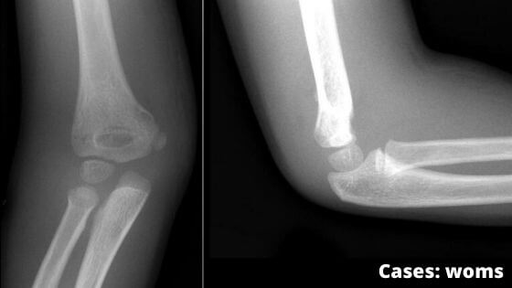
A 55-year old individual sustained a severe blow on his right flexed elbow. He developed pain and swelling in the elbow region. He was taken to an orthopedic surgeon who on examination found that the three bony points (olecranon, medial epicondyle, and lateral epicondyle) in the below region were forming an equilateral triangle. He suspected a fracture and advised the x-ray of the elbow region. The x-ray revealed the supra-epicondylar fracture of the humerus.
Questions
-
- What are the three common sites of the fracture of the shaft of the humerus? Name the nerves related to these sites?
- Why the triangular relation of three bony joints is not disturbed in the supracondylar fracture of the humerus
- Which is the most commonly injured nerve in the supracondylar fracture of the humerus?
- On clinical examination, how will you differentiate the supracondylar fracture of the humerus from the posterior dislocation of the elbow?
Answers
-
- Sites: a) surgical neck, b) mid-shaft, and C) supracondylar
- a) axillary nerve, b) radial nerve, and c) median nerve.
- In the fixed elbow, the three bony points of the elbow (olecranon, medial, and lateral epicondyles) from an equilateral triangle/ isosceles triangle. It is not disturbed in supracondylar fracture because the line of fracture lies above these points.
- Median nerve
- In the supracondylar fracture of the humerus, the triangular relationship of three bony points in the elbow region is not disturbed (vide supra), but in the posterior dislocation of the elbow, it is disturbed because of the olecranon shifts posterosuperior.
- Sites: a) surgical neck, b) mid-shaft, and C) supracondylar


